Category:Human inner ear
Jump to navigation
Jump to search
Subcategories
This category has the following 5 subcategories, out of 5 total.
Media in category "Human inner ear"
The following 119 files are in this category, out of 119 total.
-
De-Hörschnecke.ogg 1.8 s; 18 KB
-

-
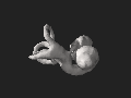 3DPX-002432 Cochlea and semicircular canals Nevit Dilmen.stl 5,120 × 2,880; 1.7 MB
-
_(14595244167).jpg/76px-A_text-book_of_the_diseases_of_the_ear_and_adjacent_organs_(1894)_(14595244167).jpg) A text-book of the diseases of the ear and adjacent organs (1894) (14595244167).jpg 1,240 × 1,948; 486 KB
A text-book of the diseases of the ear and adjacent organs (1894) (14595244167).jpg 1,240 × 1,948; 486 KB
-
_(14758743256).jpg/86px-A_text-book_of_the_diseases_of_the_ear_and_adjacent_organs_(1894)_(14758743256).jpg) A text-book of the diseases of the ear and adjacent organs (1894) (14758743256).jpg 1,312 × 1,836; 368 KB
A text-book of the diseases of the ear and adjacent organs (1894) (14758743256).jpg 1,312 × 1,836; 368 KB
-
 Akustik Basilarlen2mel2hz.jpg 779 × 540; 103 KB
-
-
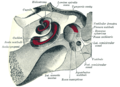
 Anatomie des Innenohrs.png 2,500 × 2,500; 3.43 MB
-
 Anatomy of the ear, John Cunningham Saunders, 1806 Wellcome L0035338.jpg 2,900 × 4,206; 3.82 MB
-
.png/120px-Balance_Disorder_Illustration_A_(hy).png) Balance Disorder Illustration A (hy).png 406 × 338; 81 KB
Balance Disorder Illustration A (hy).png 406 × 338; 81 KB
-
 Balance Disorder Illustration A zh.png 250 × 208; 22 KB
-
 Balance Disorder Illustration A-ar.png 250 × 208; 24 KB
-
 Balance Disorder Illustration A-ru.png 250 × 208; 12 KB
-
 Balance Disorder Illustration A.png 250 × 208; 15 KB
-
 Balance Disorder Illustration B.png 275 × 389; 16 KB
-
 Balance Disorder Illustration C.png 292 × 531; 12 KB
-
 Balancing act at 2012 Hong Kong Bodybuilding Championship.jpg 4,272 × 2,848; 5.77 MB
-
.JPG/90px-Basel_2012-10-05_Batch_2_(21).JPG) Basel 2012-10-05 Batch 2 (21).JPG 2,736 × 3,648; 3.06 MB
Basel 2012-10-05 Batch 2 (21).JPG 2,736 × 3,648; 3.06 MB
-
.JPG/90px-Basel_2012-10-05_Batch_2_(22).JPG) Basel 2012-10-05 Batch 2 (22).JPG 2,736 × 3,648; 3.12 MB
Basel 2012-10-05 Batch 2 (22).JPG 2,736 × 3,648; 3.12 MB
-
.JPG/120px-Basel_2012-10-05_Batch_2_(23).JPG) Basel 2012-10-05 Batch 2 (23).JPG 3,648 × 2,736; 3.61 MB
Basel 2012-10-05 Batch 2 (23).JPG 3,648 × 2,736; 3.61 MB
-
 Bigotolith.jpg 700 × 292; 105 KB
-
 BilateralVestibularHypofunction NL.png 808 × 1,499; 210 KB
-
 Blausen 0244 CochlearImplant 01.png 1,500 × 1,500; 1.45 MB
-
 Blausen 0329 EarAnatomy InternalEar EUSKARATUA.png 2,500 × 2,500; 2.67 MB
-
 Blausen 0329 EarAnatomy InternalEar ku.png 909 × 779; 376 KB
-
 Blausen 0329 EarAnatomy InternalEar-es.png 2,500 × 2,500; 3.9 MB
-
 Blausen 0329 EarAnatomy InternalEar.png 2,500 × 2,500; 3.02 MB
-
 Bony labyrinth finnish.png 250 × 208; 22 KB
-
 Bony labyrinth-ru.jpg 250 × 208; 10 KB
-
 Bony labyrinth.png 250 × 208; 12 KB
-
 Bourgery & Jacob-o3b.jpg 2,146 × 1,988; 2.12 MB
-
 BPPVvertigo NL.png 791 × 1,567; 217 KB
-
 Britannica Ear 2.jpg 276 × 161; 13 KB
-
 Canaux osseux.es.png 2,224 × 1,576; 1.07 MB
-
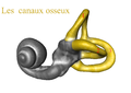 Canaux osseux.png 2,224 × 1,576; 971 KB
-
 Choclea and vestibular system 160009.jpg 2,560 × 1,920; 1.79 MB
-
 Cochlea and semicirculars MRI 115700 MAIP cr.png 480 × 404; 120 KB
-
 Cochlea and semicirculars MRI 115700 MAIP.png 490 × 506; 263 KB
-
 Cochlea and vestibular system.gif 480 × 480; 4.71 MB
-
 Cochlée.png 2,224 × 1,576; 948 KB
-
_2.jpg/120px-Cranium_-_os_petrosum_(basis_cranii)_2.jpg) Cranium - os petrosum (basis cranii) 2.jpg 4,608 × 3,456; 3.22 MB
Cranium - os petrosum (basis cranii) 2.jpg 4,608 × 3,456; 3.22 MB
-
.jpg/120px-Cranium_-_os_temporale_(os_petrosum_detail).jpg) Cranium - os temporale (os petrosum detail).jpg 4,608 × 3,456; 4.45 MB
Cranium - os temporale (os petrosum detail).jpg 4,608 × 3,456; 4.45 MB
-
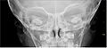
 Cross-section of a skull including detail of inner ear. Penc Wellcome V0008223.jpg 2,900 × 2,442; 2.46 MB
-
 De structura fenestrae rotuundae Wellcome M0017235.jpg 4,292 × 2,460; 4.06 MB
-
 Demonstration of Bárány chair.jpg 1,664 × 1,023; 921 KB
-
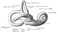
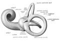
 Ear labyrinth.jpg 1,200 × 980; 680 KB
-
-
 FE Simulations Saccule.jpg 366 × 484; 68 KB
-
 Figure 36 04 04 esp.jpg 544 × 432; 107 KB
-
 Figure 36 04 04.jpg 544 × 432; 133 KB
-
 Gleichgewichtsorgan.jpg 384 × 480; 28 KB
-
 Gray900.png 887 × 1,059; 759 KB
-
 Gray901.png 873 × 1,101; 889 KB
-
 Gray920-labels.png 500 × 365; 110 KB
-
 Gray920.png 500 × 365; 46 KB
-
 Gray921 ja.png 600 × 469; 255 KB
-
 Gray921.png 600 × 469; 60 KB
-
 Gray922.png 500 × 463; 56 KB
-
 Gray923.png 600 × 434; 61 KB
-
 Gray924.png 500 × 384; 17 KB
-
 Gray926.png 600 × 417; 47 KB
-
 Gray933.png 400 × 288; 56 KB
-
_(1901).jpg/120px-Horizontal_section_through_the_right_cochlea_(A_Text-Book_on_Diseases_of_the_Ear%2C_Nose_and_Throat)_(1901).jpg)
-
In-Vivo-Analysis-of-Lrig-Genes-Reveals-Redundant-and-Independent-Functions-in-the-Inner-Ear-pgen.1003824.s009.ogv 20 s, 1,080 × 1,920; 4.53 MB
-
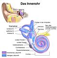 Innenohr.jpg 2,500 × 2,542; 870 KB
-
 Inner ear 1.png 460 × 367; 89 KB
-
 Inner ear Chochlea and vestibular system 115700 MAIP3 cr.png 480 × 394; 108 KB
-
 Inner ear Chochlea and vestibular system 115700 MAIP3.png 494 × 468; 208 KB
-
 Inner Ear.jpg 1,160 × 967; 74 KB
-
 Inner ear.png 460 × 367; 83 KB
-
.jpg/119px-Interior_of_the_Left_Labyrinth_of_the_Ear_in_An_academic_physiology_and_hygiene_(1903).jpg)
-
 Ird26 com044 lr Implant i örat blå.pdf 1,837 × 1,600; 353 KB
-
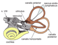
 Labyrinthe osseux Humain.png 2,224 × 1,576; 1.11 MB
-
 Malleus and Incus-Ice cream corn sign.png 507 × 507; 208 KB
-
 Medical X-Ray imaging XLC07 nevit.jpg 1,908 × 866; 261 KB
-
 Membranaotolitica.jpg 3,483 × 2,820; 1.03 MB
-
 Membranaotolitica2.jpg 3,483 × 2,820; 1.22 MB
-
_(1901).jpg/120px-Membranous_labyrinth_of_the_right_side_(A_Text-Book_on_Diseases_of_the_Ear%2C_Nose_and_Throat%2C_Plate_1)_(1901).jpg)
-
 Meniere's disease infograph VEDA.png 1,040 × 2,310; 262 KB
-
 Meyers b12 s0348a.jpg 2,048 × 1,636; 532 KB
-
_eines_11_mm_langen_menschlichen_Embryos_und_eines_29_mm_langen_menschlichen_Fetus.jpg/120px-Modelle_vom_Innenohr_(häutiges_Labyrinth)_eines_11_mm_langen_menschlichen_Embryos_und_eines_29_mm_langen_menschlichen_Fetus.jpg)
-
 Oreille Interne.png 2,518 × 2,035; 2.82 MB
-
 Otolith excitation patterns.png 931 × 462; 61 KB
-
 Otoliths w CrossSection.jpg 1,800 × 621; 208 KB
-
 OÍDO INTERNO.jpg 2,592 × 2,160; 897 KB
-
 Pendred syndrome diagram-ar.jpg 375 × 428; 31 KB
-
 Pendred syndrome diagram.jpg 375 × 428; 42 KB
-
 Place-del'Oreille-Interne-Schema.jpg 3,697 × 2,901; 2.21 MB
-
 Rocher global.jpg 13,000 × 6,500; 13.82 MB
-
 Sac,osselet.jpg 424 × 285; 42 KB
-
 SKIER2.png 750 × 607; 81 KB
-
 Sobo 1909 781.png 2,272 × 2,216; 1.99 MB
-
 Sobo 1911 772.png 2,336 × 1,876; 12.56 MB
-
 Sobo 1911 773.png 2,600 × 1,816; 13.53 MB
-
 Sobo 1911 776.png 2,408 × 1,332; 9.19 MB
-
 Sobo 1911 777.png 2,140 × 1,460; 8.95 MB
-
 Sobo 1911 778.png 2,140 × 1,144; 7.02 MB
-
 Sobo 1911 784.png 2,480 × 1,568; 11.15 MB
-
 Sobo 1911 785.png 2,436 × 1,668; 11.65 MB
-
 Sobo 1911 786.png 2,164 × 1,060; 6.57 MB
-
 Sobo 1911 787.png 1,936 × 876; 4.86 MB
-
 STS-65 fig6.png 314 × 373; 14 KB
-
 Stéréolithographie.jpg 5,008 × 4,584; 4.86 MB
-
_(14782517922).jpg/81px-Surgical_anatomy_-_a_treatise_on_human_anatomy_in_its_application_to_the_practice_of_medicine_and_surgery_(1901)_(14782517922).jpg)
-
Taeniopygia guttata song - pone.0025506.s001.oga 1.4 s; 12 KB
-
 Temporal bone1.jpg 3,168 × 2,376; 3.47 MB
-
 Temporal bone3.jpg 3,168 × 2,376; 3.32 MB
-
 Vestibular cortices and spatial cognition.jpg 569 × 850; 333 KB
-
 Vestibular neural connections.jpg 1,536 × 1,193; 339 KB
-
 Vestibular organs- canals, otolith, cochlea.jpg 527 × 430; 35 KB
-
 Vestibular pathways to cortices through the thalamus.jpg 596 × 850; 283 KB
-
 VestibularSystem la.png 300 × 229; 16 KB
-
 VestibularSystem-he.png 300 × 229; 58 KB
-
 VestibularSystem.png 300 × 229; 58 KB
-
 Vestibule.png 2,224 × 1,576; 828 KB
-
 Weightless astronauts eating.jpg 520 × 426; 224 KB
-
 Zvučni talas i mehaničke vibracije.jpg 5,120 × 1,960; 1.42 MB
-
 ヒトの耳の前庭 傾いた場合.svg 650 × 650; 131 KB
-
 ヒトの耳の前庭.svg 1,052 × 744; 179 KB

_(14595244167).jpg)
_(14758743256).jpg)


.png)


.JPG)
.JPG)
.JPG)


_2.jpg)
.jpg)


_(1901).jpg)


.jpg)


_(1901).jpg)


_eines_11_mm_langen_menschlichen_Embryos_und_eines_29_mm_langen_menschlichen_Fetus.jpg)


_(14782517922).jpg)


